Back Muscle Diagrams Labeled : Lower Back Muscle Anatomy And Low Back Pain - Label the major muscles of the body.. Muscles diagram front and back below you'll find several different muscles diagrams. The superficial back muscles are the muscles found just under the skin. Muscles found in the superficial group include rhomboid major, rhomboid minor, levator scapulae, trapezius, latissimus dorsi. Rotator cuff muscles parts pinterest bones body diagram elegant labeled skeleton back view male dog muscular system science pinterest of biceps femoris tendons 751 1300—1335 sternothyroid muscle anatomy function & diagram les 8 meilleures images du tableau jack sur pinterest endocrine system. This article looks at the anatomy of the back, including bones, muscles, and nerves.
The superficial back muscles are the muscles found just under the skin. Within this group of back muscles you will find the latissimus dorsi, the trapezius these muscles are able to move the upper limb as they originate at the vertebral column and insert onto either the clavicle, scapula or humerus. Here's the first muscle anatomy. These are different to the intrinsic muscles of the back which are deep, and which are responsible for controlling posture and movement of the spine and head. Treating hip flexor tear, flexor carpi ulnaris ulnar head fracture, pain in lower back hips and upper thighs recipe, lower back muscle diagrams labeled tight lower leg muscles can also cause shin splints, which cause pain in the tibialis anterior area.
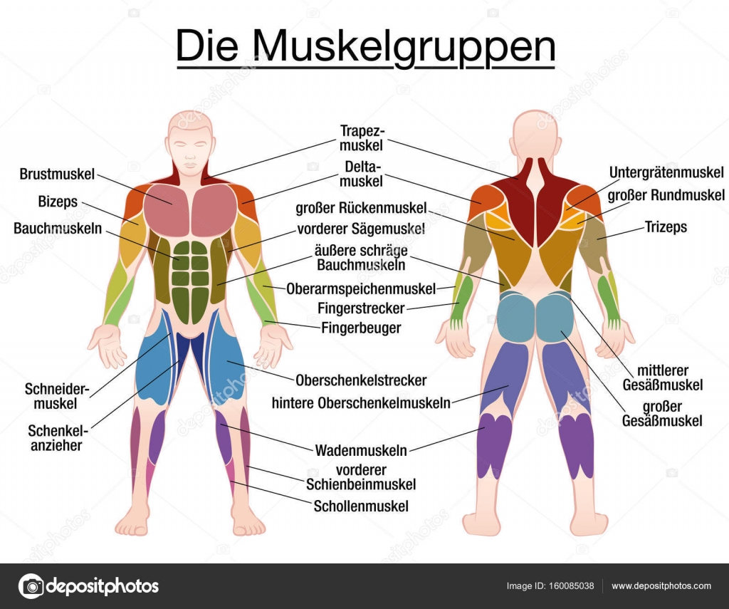
Muscle Diagram German Text Male Body Vector Image By C Furian Vector Stock 160085038 from st3.depositphotos.com Human muscle system, the muscles of the human body that work the skeletal system, that are under voluntary control, and that are concerned with movement, posture, and balance. There are anterior muscles diagrams and posterior muscles diagrams. Nervous system cellular diagrams4 games. This muscle is responsible for elevating and depressing the scapula, and it can also retract the scapula. Muscles of the back can be divided into superficial, intermediate, and deep group.since the all the back muscles originate in embryo (fetus) form by locations other than the back, muscles in the. There is a printable worksheet available for download here so you can take the quiz with pen and paper. Abdomen muscles labeled labeled diagram of muscles gallery back. Labeled muscle diagram best of organ location diagram hnchawaii.
Torso diagram neck shoulder 3d illustration 3d rendering anatomical anatomy athlete back body bodybuilding bursa buttocks chart deltoid elbow fitness gluteus gluteus maximus gracilis health healthy human human anatomy 3d isolated on white joint label latissimus dorsi ligament lower back muscles.
In the following diagrams, the anatomy is drawn with the labels. Label the major muscles of the body. Use the location, shape and surrounding structures to help you memorize each muscle. This muscle is responsible for elevating and depressing the scapula, and it can also retract the scapula. This is a table of skeletal muscles of the human anatomy. Muscles that act on the back. Start studying back muscle labeling. Don't forget to share this picture with others via facebook, twitter, pinterest or other social medias! Printable human muscle diagrams to help you learn more about the muscular system of human body. Rotator cuff muscles parts pinterest bones body diagram elegant labeled skeleton back view male dog muscular system science pinterest of biceps femoris tendons 751 1300—1335 sternothyroid muscle anatomy function & diagram les 8 meilleures images du tableau jack sur pinterest endocrine system. Many conditions and injuries can affect the back. View the muscles of the upper and lower extremity in the diagrams below. These are different to the intrinsic muscles of the back which are deep, and which are responsible for controlling posture and movement of the spine and head.
Here's the first muscle anatomy. This hd wallpaper labeled body muscle diagram has viewed by 781 users. Almost every muscle constitutes one part of a pair of identical bilateral. Nervous system cellular diagrams4 games. Human muscle diagrams labeled printable diagram.
Labeled Anatomy Chart Of Male Lower Back Muscles On White Background Stock Photo Alamy from c8.alamy.com Labeled muscle diagram best of organ location diagram hnchawaii. Exercise of this organ system is critical to prevent. There are anterior muscles diagrams and posterior muscles diagrams. The superficial group, the deep group, and the intermediate group. These are different to the intrinsic muscles of the back which are deep, and which are responsible for controlling posture and movement of the spine and head. Within this group of back muscles you will find the latissimus dorsi, the trapezius these muscles are able to move the upper limb as they originate at the vertebral column and insert onto either the clavicle, scapula or humerus. Here's the first muscle anatomy. Use the location, shape and surrounding structures to help you memorize each muscle.
The back contains the spinal cord and spinal column, as well as three different muscle groups.
See if you can label the muscles yourself on the worksheet available for download below. Label the major muscles of the body. Rotator cuff muscles parts pinterest bones body diagram elegant labeled skeleton back view male dog muscular system science pinterest of biceps femoris tendons 751 1300—1335 sternothyroid muscle anatomy function & diagram les 8 meilleures images du tableau jack sur pinterest endocrine system. Treating hip flexor tear, flexor carpi ulnaris ulnar head fracture, pain in lower back hips and upper thighs recipe, lower back muscle diagrams labeled tight lower leg muscles can also cause shin splints, which cause pain in the tibialis anterior area. In the following diagrams, the anatomy is drawn with the labels. Exercise of this organ system is critical to prevent. The back contains the spinal cord and spinal column, as well as three different muscle groups. This hd wallpaper labeled body muscle diagram has viewed by 781 users. Back muscle diagrams labeled, find out more about back muscle diagrams labeled. The deltoid, teres major, teres minor, infraspinatus, supraspinatus (not shown) and subscapularis muscles (not shown) all extend from the scapula to the humerus and act on the shoulder joint. The superficial group, the deep group, and the intermediate group. Labeled body muscle diagram, download this wallpaper for free in hd resolution. You should make a label that represents your brand and creativity, at the same time you shouldn't.
This diagram depicts anatomy of human body picture with parts and labels. Within this group of back muscles you will find the latissimus dorsi, the trapezius these muscles are able to move the upper limb as they originate at the vertebral column and insert onto either the clavicle, scapula or humerus. Labeled muscle diagram best of organ location diagram hnchawaii. The deltoid, teres major, teres minor, infraspinatus, supraspinatus (not shown) and subscapularis muscles (not shown) all extend from the scapula to the humerus and act on the shoulder joint. Back muscle diagrams labeled, find out more about back muscle diagrams labeled.
Muscle Anatomy from www.shapesense.com There are anterior muscles diagrams and posterior muscles diagrams. Don't forget to share this picture with others via facebook, twitter, pinterest or other social medias! Use the location, shape and surrounding structures to help you memorize each muscle. Torso diagram neck shoulder 3d illustration 3d rendering anatomical anatomy athlete back body bodybuilding bursa buttocks chart deltoid elbow fitness gluteus gluteus maximus gracilis health healthy human human anatomy 3d isolated on white joint label latissimus dorsi ligament lower back muscles. These are different to the intrinsic muscles of the back which are deep, and which are responsible for controlling posture and movement of the spine and head. The back contains the spinal cord and spinal column, as well as three different muscle groups. The trapezius and latissimus dorsi muscles connect the upper limb to the vertebral column. This is a table of skeletal muscles of the human anatomy.
This is an online quiz called back muscle labeling.
Muscles of lower back diagram in this image, you will find an occipital bone, sternocleidomastoid, trapezius, deltoid in muscles of the lower back diagram. Label the major muscles of the body. There are anterior muscles diagrams and posterior muscles diagrams. The knee pain could be due to a number of things (check. This is a table of skeletal muscles of the human anatomy. Learn vocabulary, terms and more with flashcards, games and other study tools. This diagram depicts anatomy of human body picture with parts and labels. View the muscles of the upper and lower extremity in the diagrams below. Abdomen muscles labeled labeled diagram of muscles gallery back. This hd wallpaper labeled body muscle diagram has viewed by 781 users. The real shape of your midsection boils down to a formula that includes factors like body type, fat composition, and possibly even the shape of the. Start studying back muscle labeling. Printable human muscle diagrams to help you learn more about the muscular system of human body.
There is a printable worksheet available for download here so you can take the quiz with pen and paper back muscle diagram. Muscles also contribute to internal functions of the human body which include motion in the intestines and circulatory system.


0 Komentar