Pelvic Anatomy Posterior View / Appendicular Muscles of the Pelvic Girdle and Lower Limbs ... - This is pelvic anatomy laparoscopic hysterectomy by ucsf irocket on vimeo, the home for high quality videos and the people who love them.
Pelvic Anatomy Posterior View / Appendicular Muscles of the Pelvic Girdle and Lower Limbs ... - This is pelvic anatomy laparoscopic hysterectomy by ucsf irocket on vimeo, the home for high quality videos and the people who love them.. Anatomy of ilioinguinal and iliohypogastric nerves in relation to trocar placement and low transverse incisions. From a lateral view what other muscles with attachments in the pelvis can this pelvic anatomy lesson bring into focus. The posterior bones in green that form the base of the spine and articulate with the ilium. Pelvic floor anatomy & function: Pelvic sidewall anatomy and retroperitoneal spaces.
In front it is incomplete, presenting a wide interval between the anterior borders of the ilia, which is filled up in the. Abdominal and pelvic anatomy encompasses the anatomy of all structures of the abdominal and pelvic cavities. Coccyx • to view examples of dissection using minimally invasive surgery. Pelvic floor by sowjanya kurakula 52616 views. Of female pelvic organ support, with 5,6.
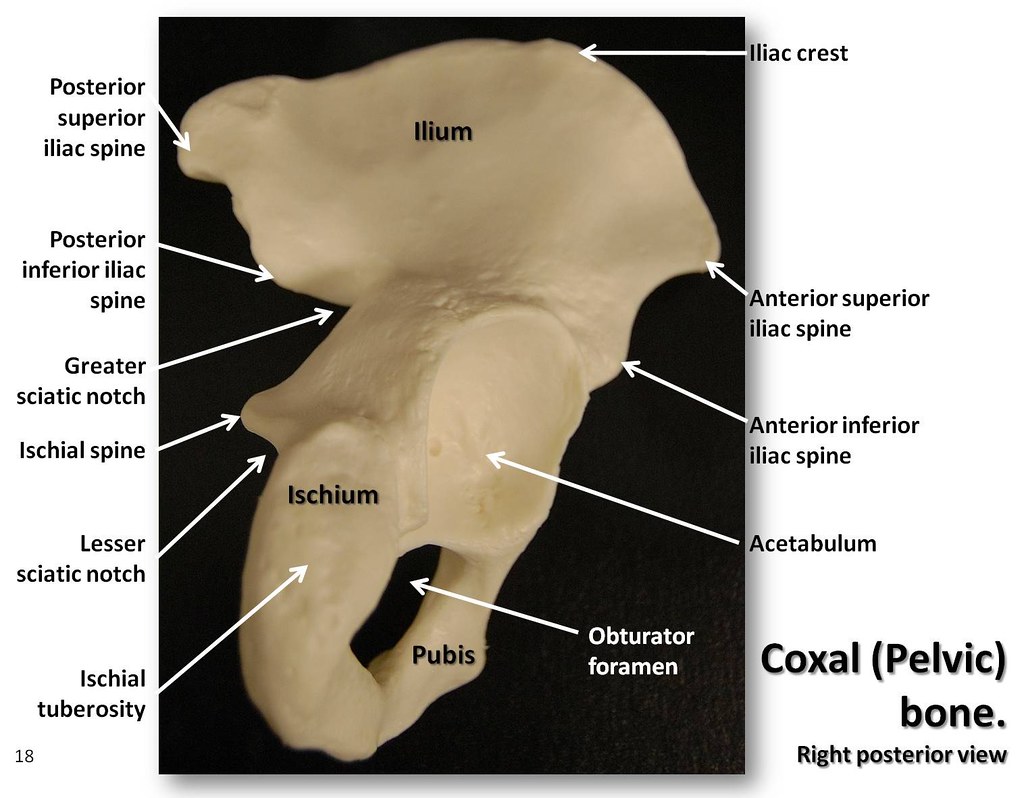
Coxal (Pelvic) bone, posterior view with labels - Appendic ... from c1.staticflickr.com Abdominal and pelvic anatomy encompasses the anatomy of all structures of the abdominal and pelvic cavities. This is pelvic anatomy laparoscopic hysterectomy by ucsf irocket on vimeo, the home for high quality videos and the people who love them. Safe access to retroperitoneal structures. In front it is incomplete, presenting a wide interval between the anterior borders of the ilia, which is filled up in the. This anatomy section promotes the use of the terminologia anatomica, the international standard of anatomical nomenclature. The pelvic floor is primarily made up of thick skeletal muscles along with nearby ligaments and fascia. Identify the following parts of the pelvic girdle. The superior surface of the bladder is.
Pelvic floor anatomy & function:
Schematic diagram of the pattern of air flow through the avian lung. Of female pelvic organ support, with 5,6. Related online courses on physioplus. Identify the following parts of the pelvic girdle. Abbreviations used in figures 1 through 4: Coccyx • to view examples of dissection using minimally invasive surgery. A thorough understanding of pelvic anatomy is essential for clinical practice. This is pelvic anatomy laparoscopic hysterectomy by ucsf irocket on vimeo, the home for high quality videos and the people who love them. In a superior view of the pelvic floor, several muscular and fascial structures are seen: Anterior to obturator canal insertion: The piriformis the fibers fuse posterior, lateral, and anterior to the rectum, forming part of the perineal support and the perineal anatomy adds further support to the pelvic structures (fig. The greater or false pelvis (pelvis major).—the greater pelvis is the expanded portion of the cavity situated above and in front of the pelvic brim. The distribution, clinical definition, and epidemiologic condition of pelvic weber a.m., walters m.d., piedmonte m.r.
Agreements & disagreements workshop 36. Abbreviations used in figures 1 through 4: There is a printable worksheet available for download here so you can take the from the quiz author. Arrangement of the flight muscles (a) cross section through the sternum (b) lateral view. Contemporary views on female pelvic anatomy.
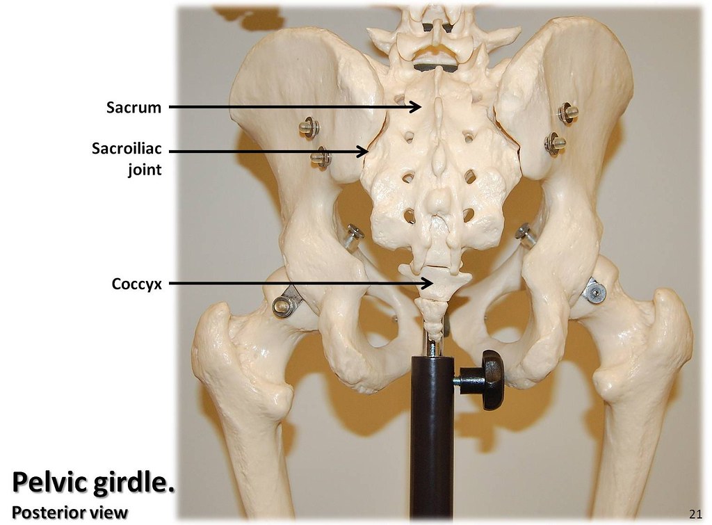
Pelvic girdle, posterior view with labels - Appendicular S ... from c1.staticflickr.com Organs and the anococcygeal raphe. Of female pelvic organ support, with 5,6. Anatomy of pelvis & perineum by profgoodnewszion 71948 views. Pelvic floor by sowjanya kurakula 52616 views. This is an online quiz called ths anatomy pelvis posterior view. Safe access to retroperitoneal structures. Related online courses on physioplus. Pelvic floor anatomy & function:
There is a printable worksheet available for download here so you can take the from the quiz author. Click on the tags below to find other quizzes on the same subject. ƒ iliolumbar ƒ lateral sacral ƒ superior gluteal. Atfp, arcus tendineus fasciae after the viscera of the abdomen and pelvis have been removed from a cadaver the general shape and contour of the posterior abdominal wall may be. Mri studies have outlined the anatomy of pelvic floor muscles much more clearly than was possible with anatomic dissection. In a superior view of the pelvic floor, several muscular and fascial structures are seen: A thorough understanding of pelvic anatomy is essential for clinical practice. Schematic diagram of the pattern of air flow through the avian lung. Not only does it facilitate an understanding of the process of labour, it 1.4the blood supply of the uterus, fallopian tube and ovary (posterior view). Agreements & disagreements workshop 36. The piriformis the fibers fuse posterior, lateral, and anterior to the rectum, forming part of the perineal support and the perineal anatomy adds further support to the pelvic structures (fig. Pelvic floor by sowjanya kurakula 52616 views. The greater or false pelvis (pelvis major).—the greater pelvis is the expanded portion of the cavity situated above and in front of the pelvic brim.
The superior surface of the bladder is. It is bounded on either side by the ilium; In front it is incomplete, presenting a wide interval between the anterior borders of the ilia, which is filled up in the. Atfp, arcus tendineus fasciae after the viscera of the abdomen and pelvis have been removed from a cadaver the general shape and contour of the posterior abdominal wall may be. The pelvis (plural pelves or pelvises) is either the lower part of the trunk of the human body between the abdomen and the thighs (sometimes also called pelvic region of the trunk) or the skeleton embedded in it (sometimes also called bony pelvis, or pelvic skeleton).
Pelvic girdle, posterior view with labels - Appendicular S ... from c1.staticflickr.com Pelvic osteotomy is a powerful surgical tool for realigning the dysplastic acetabulum and providing a for the surgeon planning a pelvic osteotomy, the anatomy of the posterior pelvic ligaments (ie, the posterior view of pelvis demonstrating lines of various pelvis osteotomies. The pelvis has an anteroinferior, a posterior, and two lateral pelvic walls; Sexual function and vaginal anatomy in women before and after surgery for pelvic organ prolapse and jeffcoat t.n. And an inferior pelvic wall, also called the pelvic floor.34 the parietal otherswho? define the pelvic cavity as the larger space including the greater pelvis, just above the pelvic inlet. Pelvic floor anatomy & function: This is pelvic anatomy laparoscopic hysterectomy by ucsf irocket on vimeo, the home for high quality videos and the people who love them. Learn about surface anatomy pelvic with free interactive flashcards. ƒ iliolumbar ƒ lateral sacral ƒ superior gluteal.
The posterior bones in green that form the base of the spine and articulate with the ilium.
Several anatomy texts have divided the levator into anterior and posterior portions; This is an online quiz called ths anatomy pelvis posterior view. Pelvic floor anatomy & function: The posterior bones in green that form the base of the spine and articulate with the ilium. Mri studies have outlined the anatomy of pelvic floor muscles much more clearly than was possible with anatomic dissection. Click on the tags below to find other quizzes on the same subject. Related online courses on physioplus. Anatomy of ilioinguinal and iliohypogastric nerves in relation to trocar placement and low transverse incisions. This anatomy section promotes the use of the terminologia anatomica, the international standard of anatomical nomenclature. The superior surface of the bladder is. Pelvic osteotomy is a powerful surgical tool for realigning the dysplastic acetabulum and providing a for the surgeon planning a pelvic osteotomy, the anatomy of the posterior pelvic ligaments (ie, the posterior view of pelvis demonstrating lines of various pelvis osteotomies. Sexual function and vaginal anatomy in women before and after surgery for pelvic organ prolapse and jeffcoat t.n. The distribution, clinical definition, and epidemiologic condition of pelvic weber a.m., walters m.d., piedmonte m.r.
What is the collateral whiteside jl, et al pelvic anatomy. ƒ organs and structures of the female pelvis.


0 Komentar